| |
|
Curr Med Chem. 2009;16(2):157-70.
Immune-glutamatergic
dysfunction as a central mechanism of the
autism spectrum disorders.
Blaylock RL1, Strunecka A.
Abstract
Despite the great number of observations being
made concerning cellular and the molecular
dysfunctions associated with autism spectrum
disorders (ASD), the basic central mechanism of
these disorders has not been proposed in the
major scientific literature.
Our review brings evidence that
most
heterogeneous symptoms of ASD have a common set
of events closely connected with dysregulation
of glutamatergic neurotransmission in the brain
with enhancement of excitatory receptor
function by pro-inflammatory immune cytokines
as the underlying mechanism. We suggest that
environmental and
dietary excitotoxins,
mercury,
fluoride, and
aluminum
can exacerbate
the pathological and clinical problems by
worsening
excitotoxicity and
by microglial
priming.
In addition, each
has effects on cell signaling
that
can affect
neurodevelopment
and
neuronal function.
Our hypothesis opens the door to a number of new
treatment modes, including the nutritional
factors that naturally reduce
excitotoxicity and brain inflammation.
|
|
|
|
|
|
|
|
|
Blood and
brain glutamate levels in
children with autistic disorder
Tamer H. Hassana, Hadeel M. Abdelrahmana, Nelly R.
Abdel Fattahb, Nagda M. El-Masryb, Haitham M. Hashimb,
, , Khaled M. El-Gerbyc, Nermin R. Abdel Fattahd
Abstract
Despite of the great
efforts that move forward to clarify the
pathophysiologic mechanisms in autism, the cause
of this disorder, however,
remains largely unknown. There is an
increasing body of literature concerning neurochemical
contributions to the pathophysiology of autism.
We aimed to determine blood and brain levels of
glutamate in children with autistic disorder
and to correlate between them. The study included 10
children with autism and 10 age- and sex-matched healthy
controls. Blood glutamate levels were measured using
high performance liquid chromatography technique. Brain
glutamate levels were measured using proton magnetic
resonance spectroscopy.
The mean
blood
and brain glutamate levels were significantly higher in
patients
than controls (p < 0.001).
There was
highly significant
positive correlation
between blood glutamate level
and
brain glutamate levels in the four tested brain
regions (p < 0.001).
Glutamate plays an important role in the
pathogenesis of autism.
Further larger studies are required to support our
findings.
| |
| |
| |
|
Neuropharmacology. 2001 May;40(6):761-71.
Protective effects of 1 alpha,25-(OH)(2)D(3)
against the
neurotoxicity
of glutamate and
reactive oxygen species in mesencephalic culture.
Ibi M1, Sawada H, Nakanishi M, Kume T, Katsuki
H, Kaneko S, Shimohama S, Akaike A.
Abstract
This study was undertaken to determine
whether 1 alpha,25-dihydroxyvitamin D3 [1
alpha,25-(OH)(2)D(3)], an active
metabolite of vitamin D, protects
dopaminergic neurons against the
neurotoxic effects of glutamate and
dopaminergic toxins using rat
mesecephalic culture.
Brief glutamate exposure elicited cytotoxicity
in both dopaminergic and non-dopaminergic
neurons. Pretreatment, but not
co-administration, of
1
alpha,25-(OH)(2)D(3) protected both types of
neurons against the
cytotoxicity of
glutamate in a concentration- and time-dependent
manner.
The neuroprotective effect of 1
alpha,25-(OH)(2)D(3) was inhibited by the
protein synthesis inhibitor, cycloheximide. To
investigate the mechanisms of these
neuroprotective effects,
we examined the
effects of
1 alpha,25-(OH)(2)D(3) on
neurotoxicity induced by
calcium ionophore and reactive oxygen species
(ROS).
.
Pretreatment with 1 alpha,25-(OH)(2)D(3)
protected both types of neurons against
the
cytotoxicity
induced by A23187
in a concentration-dependent manner.
Furthermore, 24-h pretreatment with 1
alpha,25-(OH)(2)D(3)
concentration-dependently protected both types
of neurons from ROS-induced cytotoxicity.
A 24-h incubation with
1 alpha,25-(OH)(2)D(3) inhibited the
increase in
intracellular ROS level
following H(2)O(2) exposure.
A 24-h exposure to
1-methyl-4-phenylpyridium ion
(MPP(+))
or 6-hydroxydopamine (6-OHDA)
exerted
selective neurotoxicity on dopaminergic neurons,
and these
neurotoxic effects were
ameliorated by 1 alpha,25-(OH)(2)D(3).
These results suggest that 1
alpha,25-(OH)(2)D(3) provides protection of dopaminergic neurons
against
cytotoxicity
induced by
glutamate
and dopaminergic toxins
by facilitating
cellular functions that reduce oxidative stress.
|
| |
| |
| |
|
If Immunizations increase
neuronal Oxidative Stress & Glutamate
Levels....
1
alpha,25-(OH)(2)D(3) can provide protection at
sufficient serum levels |
|
|
|
High Risk Sub-Groups for Vaccine Injury
Each SUB-GROUP listed below
is due to
25-(OH)D deficiency |
|
|
|
Premature birth or
low
birth-weight |
|
This primate study found that, “vaccinating
premature and/or low birth weight infants may create
especially high risk [of developmental delays ].”
Premature birth is associated with many risk factors
including a higher
risk of autism .
Aluminum causes developmental delays in premature babies ,
and some vaccines contain aluminum; including the
vaccine that is given to all newborns to prevent the
sexually transmitted disease Hepatitis B. |
| |
|
|
| Sibling or parent with
Type 1
diabetes |
|
Study found a high risk in this genetically susceptible
subgroup for vaccine
induced diabetes following Hib, Whole Cell Pertussis,
MMR, and OPV vaccination.
Another study also found that the Hepatitis
B vaccine appears to be linked to type 1 diabetes |
| |
|
|
Family History of
Autoimmune disorders
|
|
Several autoimmune disorders such as rheumatoid
arthritis and type 1 diabetes have been associated with
the HLA-DR4 serotype.
HLA-DR4 has also been linked to a higher rate of vaccine
injury and autism.
Animal studies have shown that autoimmune diseases can
be triggered by viruses, aluminum hydroxide, and
thimerosal in groups that are genetically susceptible
for developing autoimmune diseases. Vaccines contain all
three of those potential triggers, and there is evidence
that autism might also be an autoimmune disease of some
type. Moreover, studies have also shown that this same
subgroup tends to be a non-responder to certain
vaccines.
For more information, see:
* Auto-immune
diseases: vaccines as a possible trigger
* Potential
genetic marker for susceptibility to vaccine-injury
* Could
Autism be an auto-immune disease
|
| |
|
|
| Vitamin D Deficiency |
|
Vitamin D deficiency “is present in up to 50% of young
adults and apparently healthy children. ”
Rickets has been on the rise in infants.
A vitamin D deficiency increases the risk of certain
autoimmune diseases such as type
1 diabetes and
multiple sclerosis.
Additionally, an association of thin bones (which can be
caused by a vitamin D deficiency) has been observed in children
with autism. |
| |
|
|
| Sibling or parent with
Asthma
|
|
The CDC found that the Hepatitis
B vaccine is associated with a 20% increased risk of
developing asthma.
And another large study found that earlier
vaccination causes asthma.
However, delaying
vaccination by just a few months appears to
significantly reduce the risk of vaccine-induced asthma. |
| |
|
|
| Poor
immune function
|
|
The NY Bureau of Immunization warns that “Virus
replication after administration of live,
attenuated-virus vaccines can be enhanced in severely
immunocompromised persons. In
general, these patients should not be administered live
vaccines ”
A perfect example of this risk is that the Rotavirus
vaccine (which is intended to prevent a virus that
causes diarrhea) is actually causing chronic diarrhea
and failure to thrive in some children: “Rotavirus
Vaccine Induced Diarrhea In Child With Severe Combined
Immune Deficiency ”
The NY Bureau of Immunization also states that although
“Killed or inactivated vaccines do not represent a
danger to immunocompromised” people, their
antibody production is “suboptimal ”. |
| |
|
|
|
Mitochondrial Disease
&
Function |
|
Hannah Poling, who had an underlying mitochondrial
disorder, regressed into autism after receiving 9
vaccines in one day.
On March 29, Gerberding told CNN’s Dr. Sanjay Gupta
that, “If a child was immunized, got a fever, had other
complications from the vaccines, and (is) pre-disposed
with the mitochondrial disorder, it can certainly set
off some damage…. Some of these symptoms can be symptoms
that have characteristics of autism. I think we have to
have an open mind about this.”
http://www.ageofautism.com/2008/06/hannah-poling-r.html?cid=119382874 |
|
|
|
|
|
|
|
| RISK OF VACCINE INJURY FOR
DEFICIENT INFANTS |
| "Vaccine Injury Risk"
dependent on
Mother's 25-OH-VitD Serum
levels
during neurodevelopment |
* Each Mothers Vitamin D blood levels are passed on
to the fetus.
If the
mothers levels are LOW ...... so are the childs! |
| |
What is the optimal 25-hydroxy Vitamin D serum blood
levels
to prevent the fetus defects due to deficiency?
OPTIMAL RANGE
* For pregnant women
80ng
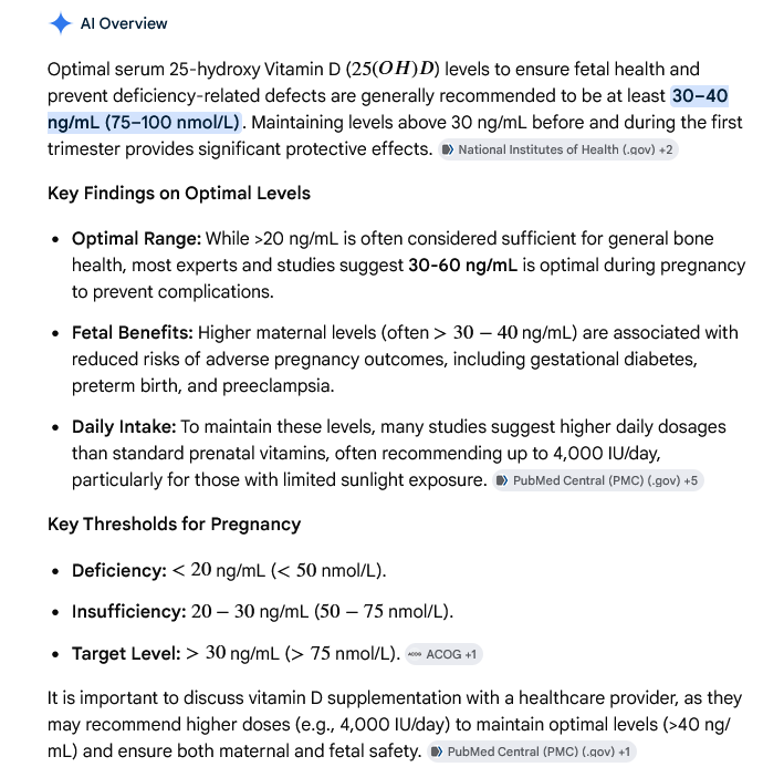 |
| |
| |
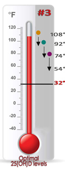
| BRAIN
FREEZES UP AT 32°
= AUTISM INITIATION |
| |
As noted above, when a
Mother's
25-OH-VitD Serum levels are TOO LOW
the fetus is at higher risk of suffering
life long deficits, like Autism. |
| |
| WATER AND ICE ANALOGY |
Water is liquid ...... until it
reaches the temperature of
32°.
When the temperature goes below 32°,
that liquid FREEZES and you now have
ICE.
LOW VITAMIN D THRESHOLD
Research indicates these AUTISTIC CHILDRENS
brains/nuerons have crossed over a threshold that
has switched off their nueral pathways,
due to the downregulation of Autophagy!
AS SHOWN BELOW
With the 32°threshold
as an anology, the mother with the
OPTIMAL Vit D serum
levels
gave her child ENOUGH of
this critical hormone to prevent the
"shut down" of the childs
autophagic pathway, due
to the 3 vaccine induced neurological
insults (shots). |
| |
| |
| Mother #1 |
| Mother is
Very
Deficient in serum
25-OH-VitD |
| * Mother passes
severe deficiency to child |
| |
| Child is at
great risk of Autism |
1st round
of immunization shots
increases oxidative stress/disease
initiation |
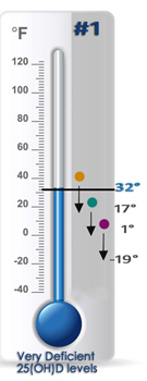
|
1st
Immunization panel sent child
below 32° |
 |
1st Vaccine Shot
- 16°
drop |
 |
2nd
Vaccine Shot - 18°
drop |
 |
3rd
Vaccine Shot - 20°
drop |
|
| Mother #2 |
|
Mother is
Deficient
in serum 25-OH-VitD |
| * Mother passes
deficiency to child |
| |
|
Child is at moderate risk of
Autism |
3rd round of immunization shots
eclipses oxidative stress
threshold/disease initiation |
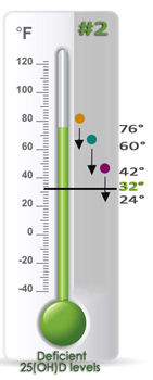
|
3rd
Immunization
panel
sent child
below 32° |
|
|
1st Vaccine Shot
- 16°
drop |
|
|
2nd
Vaccine Shot - 18°
drop |
|
|
3rd
Vaccine Shot - 20°
drop |
|
| Mother #3 |
|
Mother has
Optimal
serum 25-OH-VitD levels |
| * Mother passes
optimal levels to child |
| |
|
Child is not at risk of
developing Autism |
Immunizations
do not initiate pathology
due to sufficient 25-OH-VitD levels |
.
|
Immunization panels didn't send child
below 32° |
|
|
1st Vaccine Shot
- 16°
drop |
|
|
2nd
Vaccine Shot - 18°
drop |
|
|
3rd
Vaccine Shot - 20°
drop |
|
|
|